
Glomerular Disease Primer: The Normal Kidney
Kidney Anatomy and Physiology
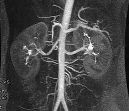
The kidneys are paired structures that lie in the retroperitoneum (behind the abdominal organs, including the liver, spleen and intestines). In an adult, each kidney is about 10-12 cm high. The kidneys get their blood supply via the renal arteries, shown below in the arteriogram as branches of the abdominal aorta (the major artery that runs along the spine).
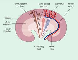
The kidney has an outer rim (cortex) and an inner core (medulla).
Each kidney contains approximately 500,000 glomeruli, which are small balls of capillaries (diameter 0.1 mm) through which the blood is filtered. Glomeruli are present only in the cortex. In the healthy kidney, approximately 100 ml/min is filtered through the glomerulus and into the space outside (urinary space). This rate is called the glomerular filtration rate (GFR). Thus, the glomeruli filter approximately 150 liters/d. This fluid then travels along a long tubule, extending from the cortex to the medulla and back to cortex and back to medulla, where the urine empties into the renal pelvis. From there the urine enters the ureter and on to the bladder. As the urine passes along the tubule, approximately 95% of fluid and many essential salts are reabsorbed and returned to the circulation. The final urine volume is typically approximately 1-2 liters a day.
Glomerular Histology
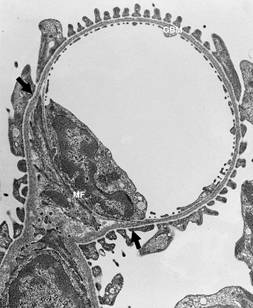
Blood flows into the glomerulus via the afferent arteriole (AA) and out via the efferent arteriole (EA). Inside the glomeruli, the blood navigates a branching set of narrow capillaries. Blood typically composed of 40% cells (white blood cells and red blood cells) and 60% plasma (straw-colored fluid, rich in proteins). Approximately 20% of the plasma that enters the glomerulus is filtered through the capillary filtration barrier and out into the urinary space (US) inside Bowman’s capsule. The remaining 80% of the plasma and all the blood cells leave the glomerulus via the efferent arteriole.
Glomeruli contain three cell types:
- endothelial cells (E) line the capillaries and provide a smooth, non-clotting surface for blood to flow smoothly over
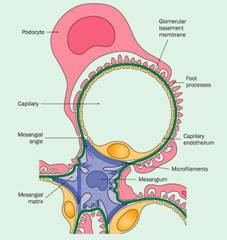
- the mesangial cells (M) are located within the central portion of the glomerulus and provide structural support
- the podocytes (PO) are located outside the capillary loops and regulate filtration of proteins
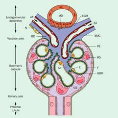
The glomerular basement membrane (GBM, shown in green) is a thin sheet of extracellular matrix protein that surrounds the capillary and provides support and a barrier.
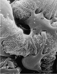
The podocytes have an especially complex structure. Each podocyte sends out primary processes (shown above in pink above and labeled as PP in the electron micrograph below) extending to the GBM. At the end of these processes, delicate foot processes (FP) rest on the GBM. Foot processes from adjacent podocytes lie next to each other and interdigitate, like the interlocking fingers of two hands. The scanning electron micrograph shown below was taken from outside the capillary loop, so that the glomerular capillary appears as a tube which is entirely covered by podocyte primary processes and foot processes.
Glomerular Filtration Barrier
The major function of the glomerulus is to allow the passage of small molecules (toxins, salts) into the urinary space while keeping most proteins in the capillary loop. Three components of the glomerulus contribute to the glomerular filtration barrier, preventing the escape of proteins into the urinary space, as shown in the electron microscope pictures below:
- endothelial cells: there are small gaps in the endothelial cell cytoplasm (shown as arrowheads) the allow most proteins to penetrate
- glomerular basement membrane (GBM, the three-part zone in the middle, with a light-dark-light pattern): this is composed of collagen and other matrix proteins and is believed to be a major barrier (perhaps the major barrier) to the passage of proteins into the urinary space
- slit-diaphragms (fine line, shown by arrows): these connect adjacent foot processes extending from the podocytes and offer a final barrier to the passage of proteins into the urinary space
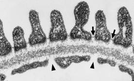
In healthy kidneys, the glomerular filtration barrier functions to allow the escape of salts and small proteins into the urinary space, while retaining larger proteins and blood cells within the glomerular capillary loop.
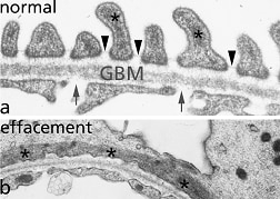
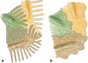
Podocyte foot process effacement. Many proteinuric diseases are characterized by loss of the glomerular slit diaphragms (left figure, top image) and the appearance of a continuous layer of podocyte cytoplasm (left figure, bottom image) This process is termed foot process effacement and is reversible, as when therapy induces a remission. The figure on the right shows schematically what is happening: the normally inter-digitating, inter-locking podocing processes have disappeared and are replaced by a simple podocyte architecture. Presumably the proteinuria occurs because plasma proteins penetrate the abnormal gaps that separate the podocytes from each other.
Acknowledgments. We wish to thank Dr. Richard Johnson and Dr. Wihelm Kriz for permission to reproduce figures.