

Ostomy Surgery Showing Diseased and Healthy Large Intestines
Description
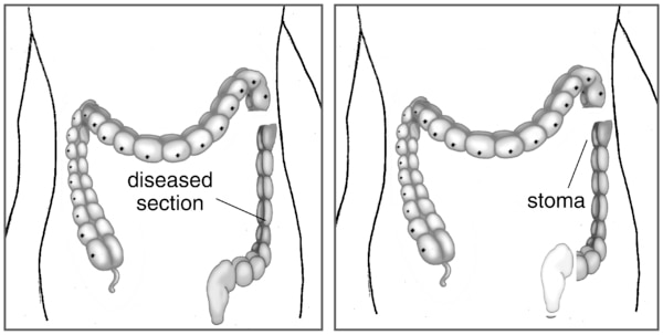
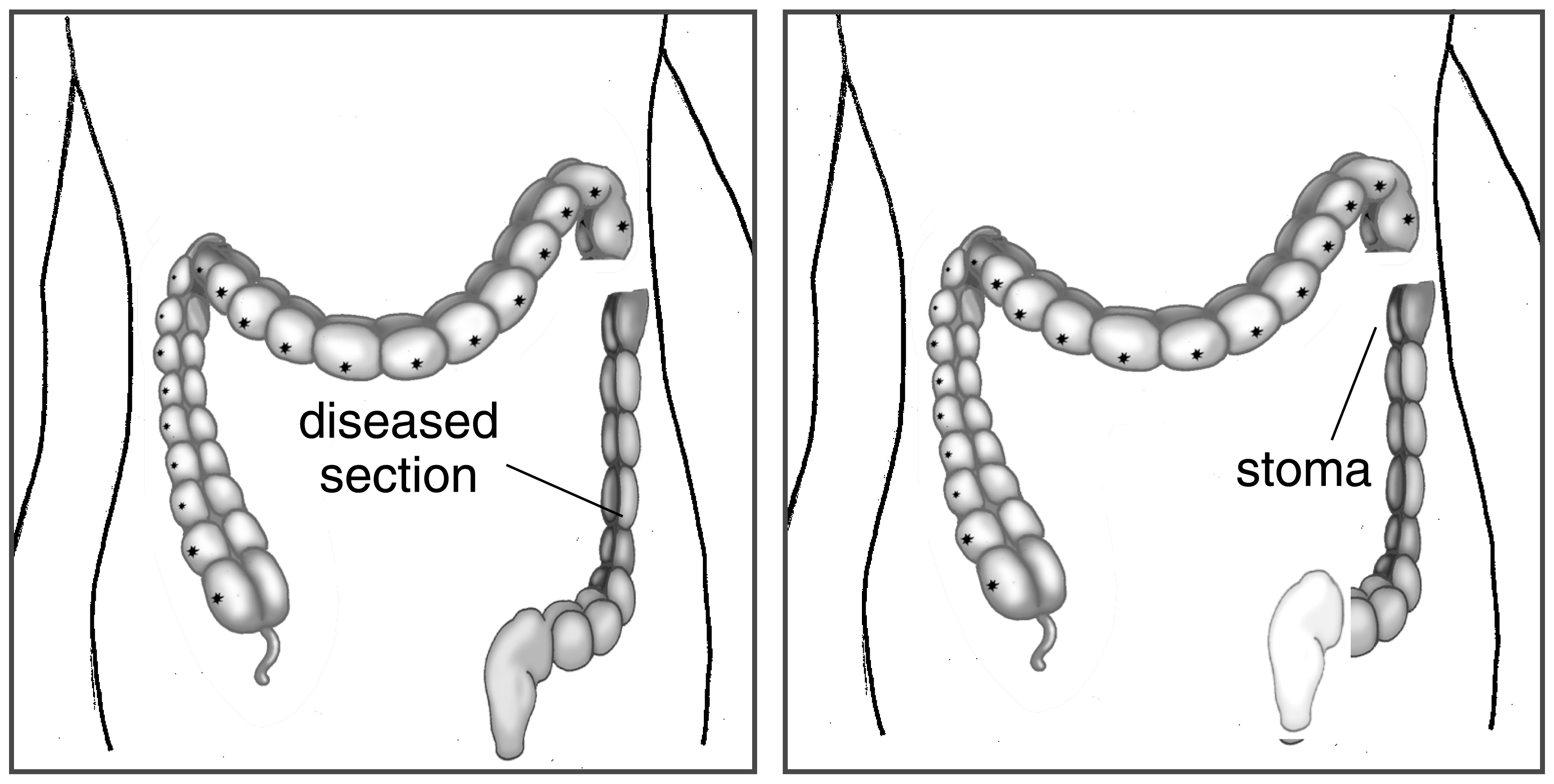
Two black and white illustrations of ostomy surgery. The first shows the large intestine with the diseased section labeled and detached from the healthy section. The second shows the healthy section attached to a stoma labeled.
Alternate Text
Two illustrations of ostomy surgery. The first shows the large intestine with the diseased section labeled and detached from the healthy section. The second shows the healthy section attached to a stoma labeled.
Caption
Step 1: The doctor takes out most of the diseased part of the intestine. Step 2: The doctor attaches the healthy part of the intestine to the stoma (a hole in the abdomen).
Diseases or Conditions
File Size
745 KB | 3440 x 1736
File Type
JPG
Share this page