

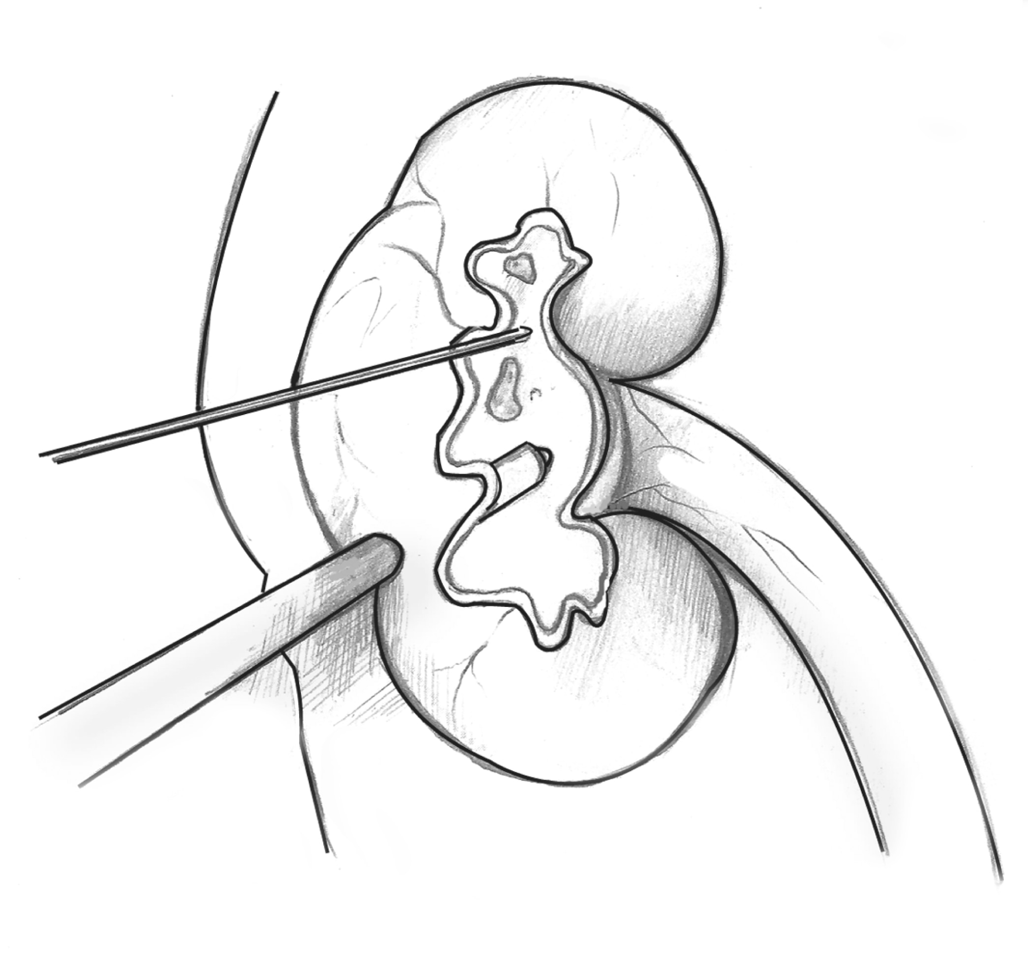
A kidney in cross-section to show an internal stone. A thin wire is inserted through the skin into the kidney to locate the stone

Description
Drawing of a kidney in cross-section to show an internal stone. A thin wire is inserted through the skin into the kidney to locate the stone. A slightly thicker probe is also inserted through the skin into the kidney to deliver sound waves that will break
Alternate Text
Drawing of a kidney in cross-section to show an internal stone. A thin wire is inserted through the skin into the kidney to locate the stone. A slightly thicker probe is also inserted through the skin into the kidney to deliver sound waves that will break
Caption
Percutaneous nephrolithotomy
Diseases or Conditions
File Size
664 KB | 2100 x 1950
File Type
JPG
Share this page