

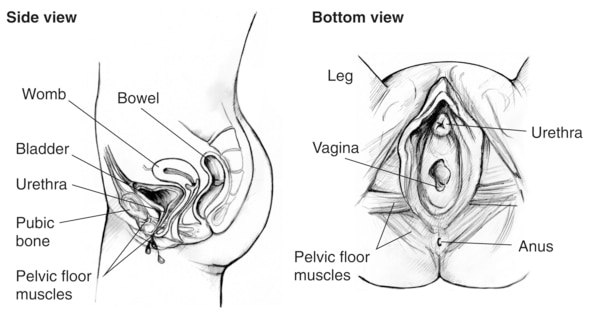
Side and bottom views of the female urinary tract
Description
Two anatomical drawings of the female urinary tract. Drawing on the left is a side view with labels pointing to the womb, bladder, urethra, public bone, and pelvic floor muscles. Drawing on the right is a bottom view with labels pointing to the leg, ureth
Alternate Text
Two anatomical drawings of the female urinary tract. Drawing on the left is a side view with labels pointing to the womb, bladder, urethra, public bone, and pelvic floor muscles. Drawing on the right is a bottom view with labels pointing to the leg, ureth
Caption
Parts of the bladder control system.
Diseases or Conditions
File Size
1.21 MB | 3000 x 1575
File Type
JPG
Share this page