

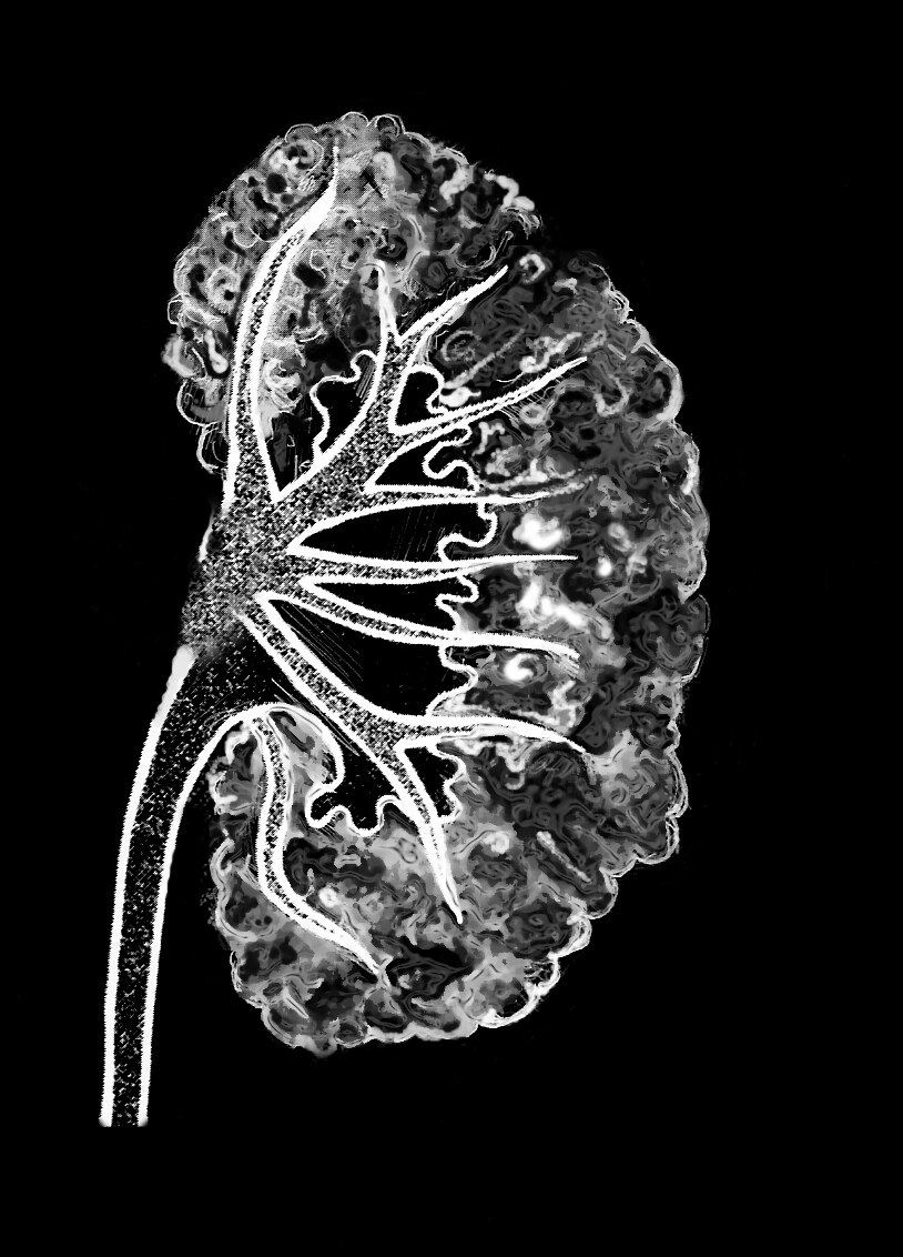
A medullary sponge kidney as seen in an intravenous pyelogram

Description
Drawing of a medullary sponge kidney as seen in an intravenous pyelogram. The background is black. The large part of the kidney appears to be porous, like a sponge.
Alternate Text
Drawing of a medullary sponge kidney as seen in an intravenous pyelogram. The background is black. The large part of the kidney appears to be porous, like a sponge.
Caption
In an intravenous pyelogram of a medullary sponge kidney, cysts appear as clusters of light.
Diseases or Conditions
File Size
368 KB | 814 x 1132
File Type
JPG
Share this page