About Our Research
Iron is an essential nutrient for almost every organism. It is required by every cell in the human body, yet it can also be a potent cellular toxin. Iron is essential because enzymes that require iron cofactors (namely, heme, iron-sulfur clusters, mononuclear and diiron centers) are involved in virtually every major metabolic process in the cell. Our laboratory focuses on the genetics and cell biology of iron uptake and utilization in eukaryotes. Previously, we have identified and characterized systems of iron transport in baker’s yeast, Saccharomyces cerevisiae. More recently, we have used the genetic tractability of yeast to focus on the intracellular trafficking and distribution of iron cofactors in yeast and mammalian cells.
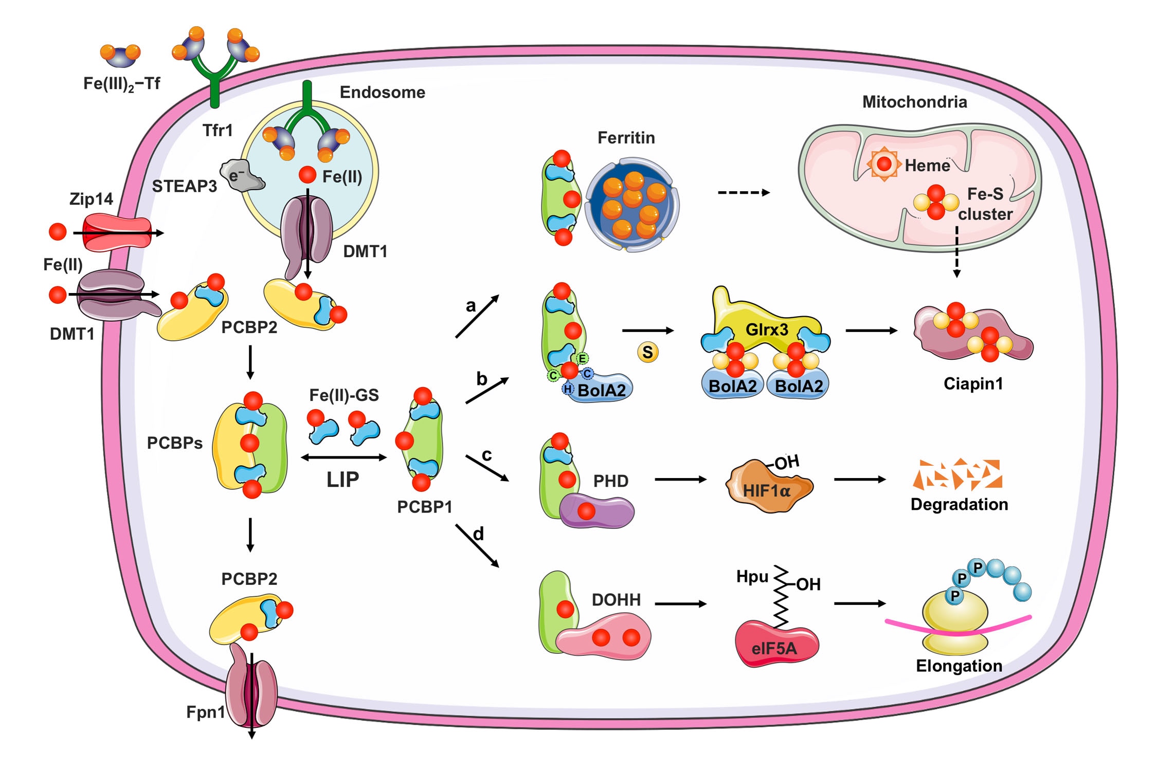
Mammalian cells express hundreds of metalloproteins. Most contain the abundant metals iron and zinc, while others contain various trace metals such as copper, manganese, molybdenum and cobalt. Although the incorporation of the appropriate metal ion(s) into cellular metalloproteins is a critical, essential process, the mechanism by which most metalloproteins receive their specific cofactor is unknown. Some proteins rely on metallochaperones: proteins that specifically bind metal ions and deliver them to target enzymes and transporters through direct protein-protein interactions.
Current Research
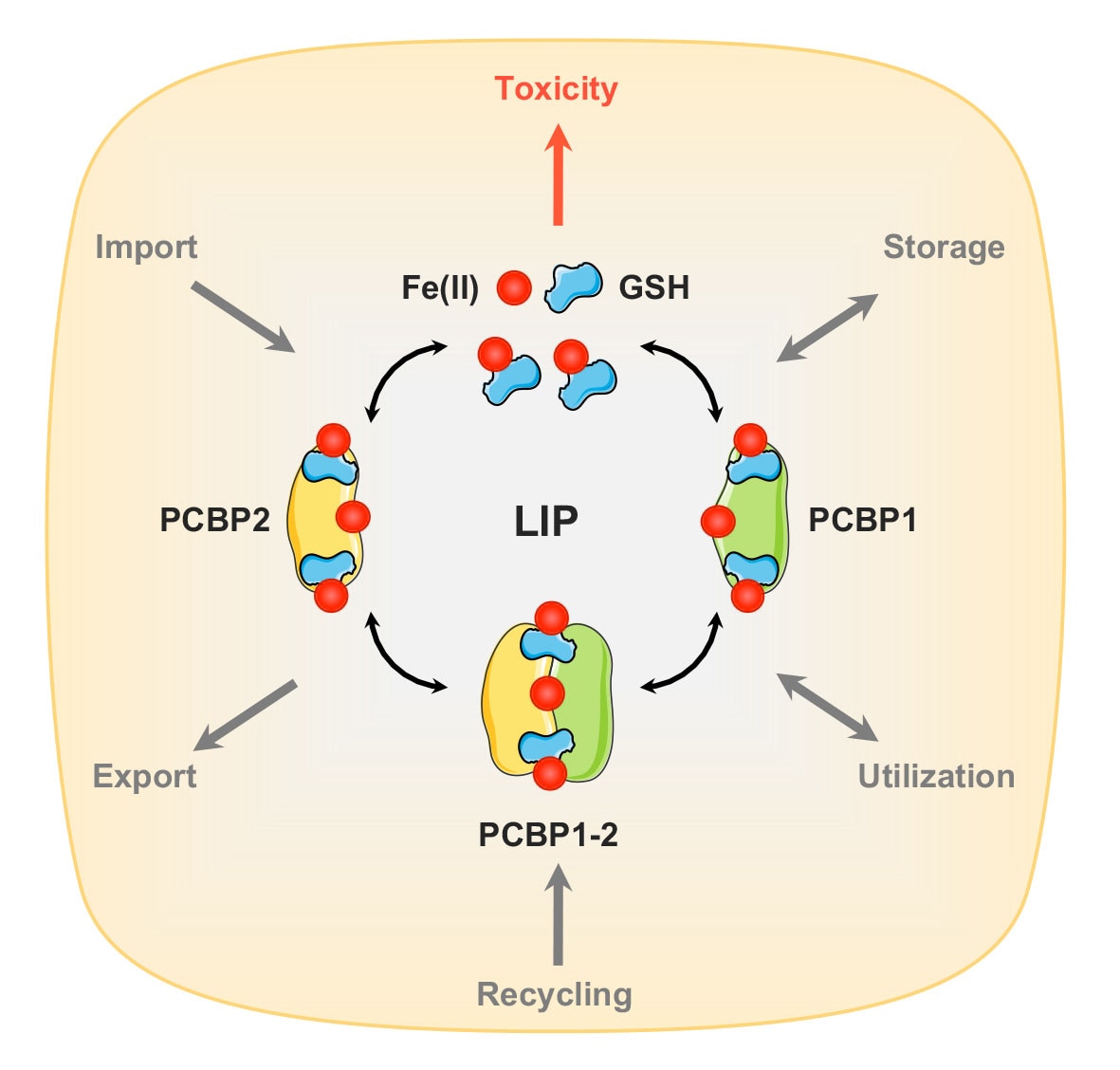
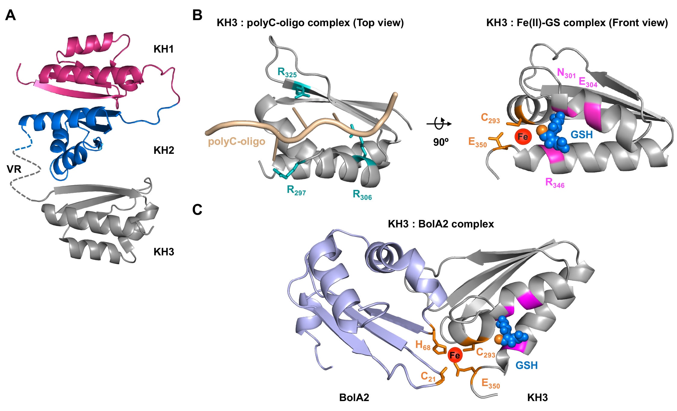
Human cells express hundreds of proteins that require metal cofactors, including dozens of proteins requiring iron in various forms. Almost nothing is known about how mono- and di-nuclear iron enzymes are metallated. PCBP1 and PCBP2 have been identified as iron chaperones: proteins that specifically bind iron and deliver it directly to target iron enzymes via a metal-mediated protein-protein interaction. Other proteins that may be involved in intracellular iron delivery are unknown. The focus of our research program is to determine i) the spectrum of iron-dependent processes controlled by PCBPs, ii) the mechanism of iron binding and transfer to target enzymes, iii) the process by which PCBPs acquire iron for delivery, iv) the biology of the PCBP system in mammalian tissues, and v) the broader roles of PCBPs in diverse functions of the cell.
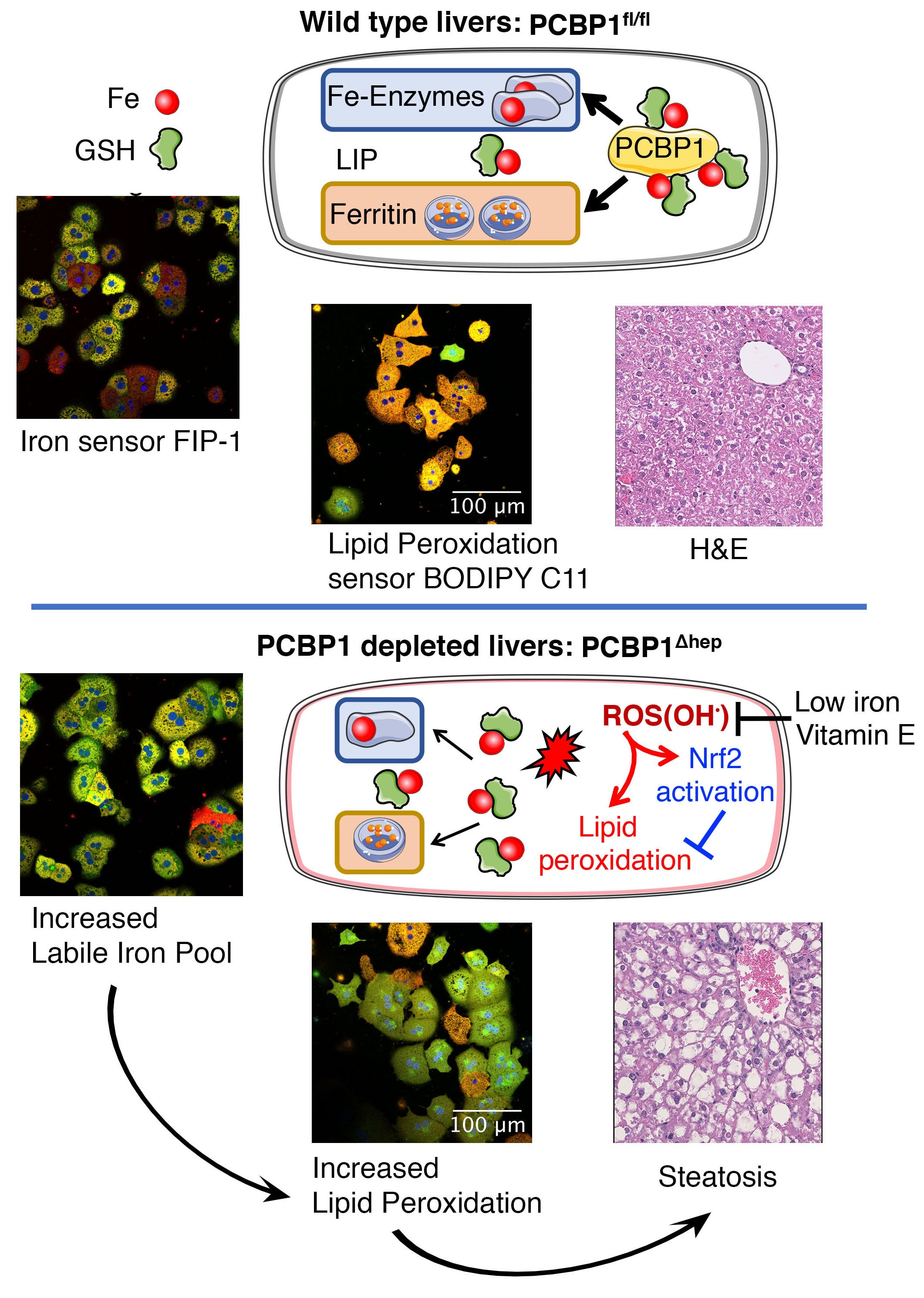
Iron deficiency continues to be the most common nutritional deficiency in the world, especially among children and women of childbearing age, where it causes anemia and impairs neurological development and function. Although the pathogenesis of anemia in iron deficiency is well understood, other manifestations of iron deficiency are not understood at the cellular or metabolic level. Iron overload is a feature of an increasing number of human diseases, including genetic disorders such as hereditary hemochromatosis, thalassemias, and Friedreich ataxia, as well as chronic inflammatory diseases of the liver, such as hepatitis C.
Research Images

Iron uptake systems release Fe(II) into the cytosol. Cytosolic Fe(II) is coordinated by reduced glutathione (GSH) to form the cytosolic LIP. Under physiological conditions, a significant proportion (>90%) of LIP may be coordinated by iron chaperones, PCBPs. Structurally, PCBPs contain three tandem K-homology domains (KH1–3) that can bind GSH coordinated Fe(II) with high affinity in a 1:1 ratio. PCBP1 and PCBP2 are the major iron chaperones that play integral roles in intracellular iron trafficking. PCBP1 and PCBP2 can bind to each other in cells, potentially transferring iron between them. Unchaperoned free Fe(II) reacts with cellular oxidants to produce cytotoxic hydroxyl radicals via the Fenton reaction.

A. A complete projected structure of PCBP1 based on K-homology (KH) domains 1 and 2 (PDB 2JZX) and domain 3 (PDB 1WVN) linked together via unstructured variable region (VR).
B. Structural comparison of PCBP1 KH3 domain in complex with RNA or iron. Left: a homology structure of poly C-oligo bound form of KH3 domain (PDB 2P2R) illustrating ligands for cytosine binding (teal sticks). Right: a ribbon structure of KH3 domain (PDB 1WVN) illustrating ligands for GSH binding (magenta) and iron binding (orange sticks), coordinating the iron (red sphere) and GSH (blue spheres, thiol in orange).
C. A model for electrostatic interaction between BolA2 (PDB 1V9J, light blue) and KH3 domain (PDB 1WVN, grey) presenting ligands for iron binding (orange sticks), coordinating the iron (red sphere) and GSH (blue spheres, thiol in orange). Models were generated using Swiss-model for homology modeling, HADDOCK 2.2 protein docking platform and PyMOL version 2.3.

Divalent metal transporter 1 (DMT1) and Zip14, located on plasma membrane or endocytic vesicles, imports non-transferrin-bound or transferrin-bound iron, respectively. Iron enters the cytosolic labile iron pool (LIP) as Fe(II) and is largely coordinated by reduced GSH and bound to iron chaperones, PCBPs. PCBP1 and PCBP2 efficiently distribute iron for storage, non-heme iron cofactor assembly, or efflux through ferroportin (Fpn1). a, PCBP1 (and to a lesser extent, PCBP2) facilitates iron sequestration into ferritin, which can be delivered to mitochondria by unknown mechanism for heme or Fe-S cluster synthesis. b, PCBP1 form a complex with cytosolic BolA2 via a bridging Fe-GS ligand. Iron provided by PCBP1-BolA2 complex can be combined with sulfur (S) compound, with the help of other cytosolic Fe-S cluster assembly proteins and facilitate the [2Fe-2S] cluster formation on BolA2-Glrx3 distribution system. c, PCBP1 delivers iron to mononuclear iron enzyme prolyl hydroxylase (PHD2), which regulates the degradation of hypoxia inducible factor 1- (HIF1). d, PCBP1 also metallates the dinuclear iron enzyme deoxyhypusine hydroxylase (DOHH), which catalyzes the hydroxylation of hypusine (Hpu) on eukaryotic translation initiation factor 5A (elF5A) to promote translation of polyproline motifs.

Reactivity of unchaperoned iron in PCBP1-depleted hepatocytes causes increased lipid peroxidation. Then, the accumulation of lipid peroxidation products damages the liver, alters lipid metabolism, and results in liver steatosis. Reactivity of iron pool and lipid peroxidation were visualized using the FIP-1 iron sensor and BODIPY C11. Morphological changes in the livers were detected by hematoxylin and eosin (H&E) staining of liver sections.