

Urinary Tract Imaging
On this page:
- What is the urinary tract?
- What does “imaging” mean?
- What symptoms could require imaging of the urinary tract?
- What are some other reasons for imaging the urinary tract?
- What steps does your health care professional take before ordering imaging tests?
- What are the imaging techniques?
- How do you prepare for an imaging test?
- What happens after your imaging test?
- Can an imaging test cause kidney damage?
- How soon will test results be available?
- Clinical Trials for Urinary Tract Imaging
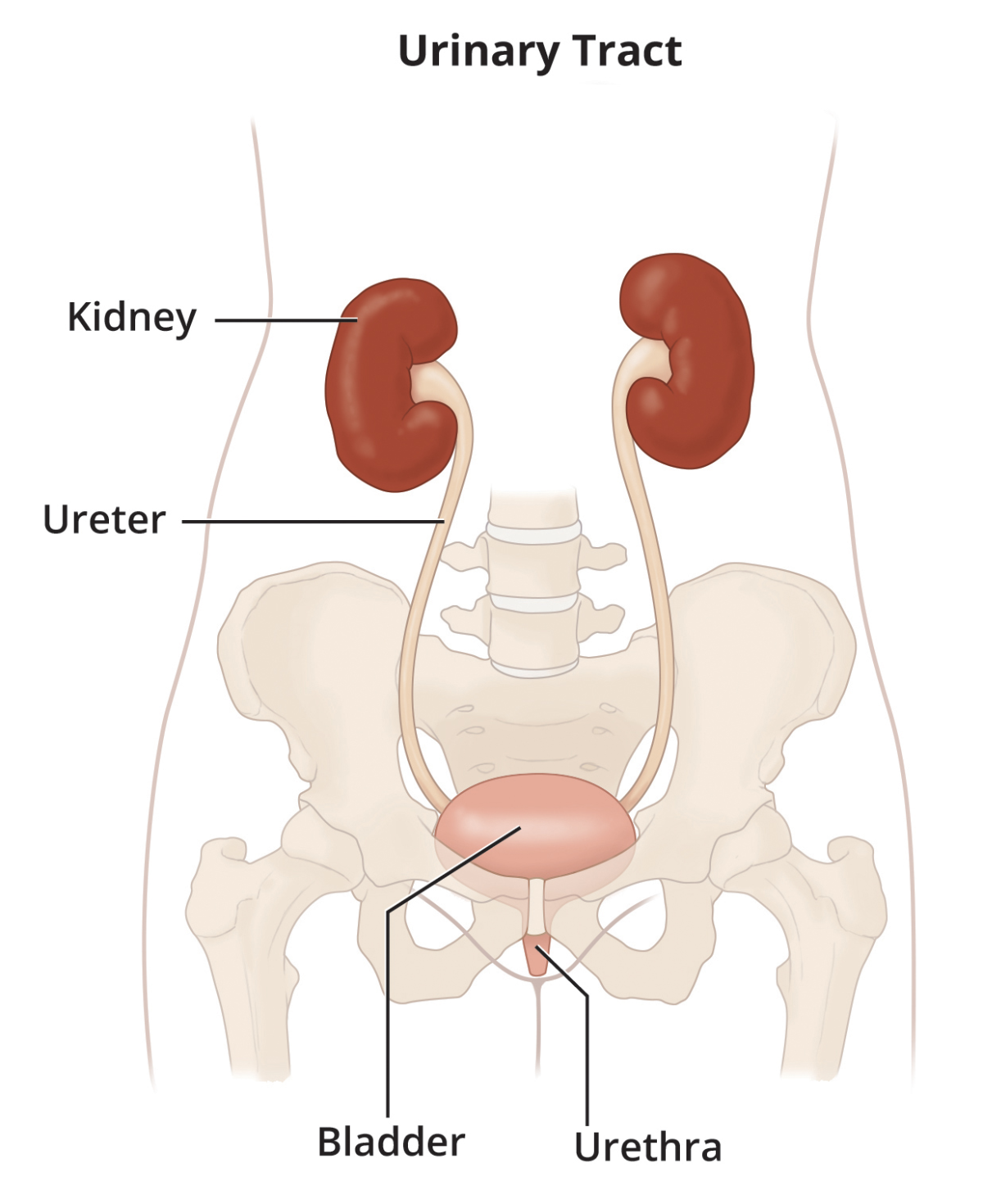
What is the urinary tract?
The urinary tract is your body’s drainage system for removing wastes and extra fluids. The urinary tract includes two kidneys, two ureters, a bladder, and a urethra.
The kidneys filter wastes and fluids to produce urine. The urine travels from the kidneys down two narrow tubes called the ureters. The urine is then stored in a hollow, muscular, balloon-shaped organ called the bladder. When the bladder empties, urine flows out of the body through a tube called the urethra at the bottom of the bladder.
What does “imaging” mean?
Imaging is a general term for techniques used to create pictures. In medicine, imaging produces pictures of bones, organs, and vessels inside the body. Imaging helps health care professionals see the cause of medical problems. Imaging techniques include
- x-rays
- ultrasounds
- magnetic resonance imaging (MRI) scans
- computed tomography (CT) scans
- radionuclide scans
What symptoms could require imaging of the urinary tract?
Imaging could be required for symptoms such as
- difficulty initiating or maintaining urination
- difficulty in emptying the bladder, known as urinary retention
- accidental leakage of urine, known as bladder control problems or urinary incontinence
- urinary frequency and urgency (day or night)
- recurrent urinary tract infections (UTIs)
- a single UTI in a susceptible or high-risk person, such as an infant
- pain in the abdomen, upper or lower back, or groin
- abdominal pain or mass, such as swelling in a specific part of the abdomen
- evidence of kidney failure
- blood in the urine, known as hematuria
- high blood pressure
What are some other reasons for imaging the urinary tract?
Your health care professional might also order urinary tract imaging to pinpoint a problem. That’s important because different urinary tract problems may share the same symptoms. For example, a urinary blockage can be caused by a kidney stone or an enlarged prostate.
Imaging can also help your health care professional identify, evaluate, follow up, and monitor problems such as
- kidney diseases
- tumors
- small bladder capacity
- backward flow of urine, known as vesicoureteral reflux (VUR)
- hydronephrosis, or urine blockage, in newborns following suspicious or abnormal imaging during the pregnancy
What steps does your health care professional take before ordering imaging tests?
Before ordering imaging tests, your health care professional will consider your general medical history, including any major illnesses or surgeries, perform a physical exam, obtain blood test results, and may ask
- about your specific urinary tract symptoms, when they began, how often they occur, and how severe they are
- if you take any prescription or over-the-counter medicines
- how much fluid you take in each day
- about your use of alcohol and caffeine
- whether you are allergic to any foods or medicines
- whether you could be pregnant, if you are a female patient
What are the imaging techniques?
Your health care professional can use several different imaging techniques, depending on factors such as your general medical history and urinary tract symptoms.
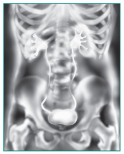
X-rays
X-rays of the urinary tract can help highlight and monitor a kidney stone or tumor that could be blocking the flow of urine and causing pain.
Conventional x-rays involve some exposure to ionizing radiation, a type of radiation strong enough to damage some cells.
Two common x-ray procedures used for urinary tract imaging include
- intravenous pyelogram (IVP) to help locate problems in the kidneys, ureters, or bladder that may be caused by urinary retention or urinary reflux
- voiding cystourethrogram (VCUG) to view images of the bladder and urethra taken while the bladder is full and during urination
Ultrasound
Ultrasound uses a hand-held device, called a transducer, that bounces safe, painless sound waves off organs to create an image of their structure. The health care professional can move the transducer at different angles to examine different organs.
This procedure is painless, poses no risk of radiation, needs no anesthesia, and allows you to return to daily tasks immediately.

Health care professionals use specific types of abdominal ultrasounds to look at different parts of the urinary tract.
- Bladder ultrasound can give information about the bladder wall, diverticula (pouches) of the bladder, bladder stones, and large tumors in the bladder.
- Kidney ultrasound can show if the kidneys are in the right place or if they have blockages, kidney stones, or tumors.
MRI scans
Magnetic resonance imaging (MRI) takes pictures of the body’s internal organs and soft tissues without using x-rays. MRI machines use radio waves and magnets to produce detailed pictures of the body’s internal organs and soft tissues. During an MRI, a special dye known as a contrast medium may be injected into the blood before the test, usually intravenously (IV) through a vein in the hand or forearm. The dye helps the radiologist see certain areas more clearly.
Magnetic resonance angiography (MRA). An MRA is a type of MRI that provides the most detailed view of kidney arteries, which are the blood vessels that supply blood to your kidneys. An MRA can also show renal artery stenosis.
Magnetic resonance urography (MRU). An MRU is a type of MRI used to evaluate patients with blood in the urine, known as hematuria. MRU is also used when following up with patients who have a history of urinary tract cancers and to identify abnormalities in patients with recurrent urinary tract infections.
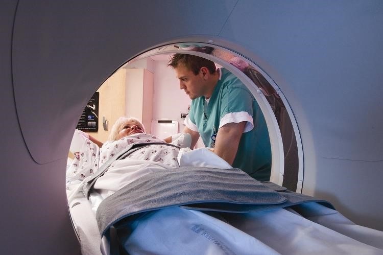
CT scans
A CT scan combines x-rays with computer technology to create three-dimensional (3-D) images. These scans can show stones in the urinary tract, as well as obstructions, infections, cysts, tumors, and traumatic injuries. Imaging for urinary stone disease can be done with low or ultra-low dose CT scans.
Radionuclide scans
A radionuclide scan, also called a nuclear scan or radioisotope scan, detects small amounts of radiation after radioactive material is injected into the blood. This scan provides information about how your kidneys function and helps health care professionals diagnose many conditions, including cancers, injuries, and infections.
Renal scan, also called kidney scan. Your health care professional might use a renal scan to check your kidneys and the urinary system. This type of exam includes injecting a small amount of radioactive material into the blood and using a special camera and computer.
There are different types of renal scans, and they can be used to check the kidneys along with other imaging methods such as ultrasound, CT scans, and MRIs. Sometimes they can provide unique information that is hard to get from other imaging procedures. A health care professional determines which method will provide the best information about your kidneys and urinary system.
PET scan. A positron emission tomography (PET) scan is a type of imaging that uses a small amount of radioactive material, a special camera, and a computer to help health care professionals see how the organs and tissues are working. PET scans are sometimes performed on combined PET/CT scanners.
How do you prepare for an imaging test?
How you prepare for an imaging test will depend on the test’s purpose and type. Your health care professional will give you instructions. Listen carefully and ask questions if you don’t understand. You might be asked to
- drink several glasses of water 2 hours before some ultrasound tests
- take a laxative for a transrectal ultrasound
- take an enema about 4 hours before a transrectal ultrasound
- talk with the technical staff about any implanted devices that may have metal parts, such as heart pacemakers, intrauterine devices (IUDs), hip replacements, implanted ports for catheters, and metallic items, such as metal plates, pins, screws, surgical staples, bullets, or shrapnel
- take a sedative before an MRI or CT scan if you feel anxious or have difficulty holding still in enclosed spaces
- discuss with your health care professional if there is any chance you may be pregnant and whether the imaging uses x-rays
- fast for 12 hours before the test
What happens after your imaging test?
After most imaging tests, you can go home and resume normal activity. Some tests that involve catheters may cause minor discomfort. Tests that include medication, dyes, or sedatives occasionally trigger allergic reactions.
Tests that may cause discomfort include
- Tests involving a catheter in the urethra. You might feel some mild discomfort from an irritated urethra for a few hours after the procedure.
- Transrectal ultrasound. You might feel some discomfort from an irritated rectum.
If you have a catheterization, your health care professional may prescribe an antibiotic for 1 or 2 days to prevent an infection. If you have any signs of infection, including pain, chills, or fever, call your health care professional immediately.
Tests that may cause an allergic reaction include
- Tests involving contrast medium. If you have a rare sign of reaction, such as hives, itching, nausea, vomiting, headache, or dizziness, call your health care professional immediately.
- Tests involving sedatives. If you have a rare sign of reaction, such as changes in breathing and heart rate, call your health care professional immediately.
Can an imaging test cause kidney damage?
A test that uses a special dye, known as contrast medium, can cause kidney damage in people with certain conditions, such as impaired kidney function or diabetes. In most people, there is no damage or damage is minimal and temporary, healing on its own within a week or so. In rare cases, contrast medium can cause lasting kidney damage. Drinking plenty of fluids before and after a CT scan with radiocontrast will dilute the dyes and help your body remove them faster, reducing the risk of kidney injury.
Kidney damage or reduced kidney function is usually diagnosed by lab testing.
How soon will test results be available?
For simple tests, such as x-rays and abdominal ultrasounds, you can discuss the results with your health care professional soon afterward. Results of other tests, such as MRIs or CT scans, may take several days to become available, and you may need a separate appointment to discuss your results.
Clinical Trials for Urinary Tract Imaging
The NIDDK conducts and supports clinical trials in many diseases and conditions, including urologic diseases. The trials look to find new ways to prevent, detect, or treat disease and improve quality of life.
What are clinical trials for urinary tract imaging?
Clinical trials—and other types of clinical studies—are part of medical research and involve people like you. When you volunteer to take part in a clinical study, you help doctors and researchers learn more about disease and improve health care for people in the future.
Researchers are studying many aspects of imaging procedures to diagnose and treat different urinary tract diseases. For example, researchers are studying
- ultrasounds for patients with kidney stones and ureter stones
- MRI and CT scans for diagnosing patients with kidney tumors
- contrast exposure tests using CT scans for patients with acute kidney injury
Find out if clinical research trials are right for you.
Watch a video of NIDDK Director Dr. Griffin P. Rodgers explaining the importance of participating in clinical trials.
What clinical trials for urinary tract imaging are looking for participants?
You can view a filtered list of clinical trials on urinary tract imaging that are open and recruiting at ClinicalTrials.gov. You can expand or narrow the list to include clinical studies from industry, universities, and individuals; however, the National Institutes of Health does not review these research studies and cannot ensure they are safe. Always talk with your health care professional before you participate in clinical research.
This content is provided as a service of the National Institute of Diabetes and Digestive and Kidney Diseases
(NIDDK), part of the National Institutes of Health. NIDDK translates and disseminates research findings to increase knowledge and understanding about health and disease among patients, health professionals, and the public. Content produced by NIDDK is carefully reviewed by NIDDK scientists and other experts.
The NIDDK would like to thank:
Ann E. Stapleton, M.D., FACP, FIDSA University of Washington School of Medicine